The ulna bone may also be broken. While bone wrist fractures are more severe than others the most common sign of a break in the distal radius is intense pain.

X Ray Fraktur Tulang Fraktur Jari Jari Distal Lengan Bahu Lengan Tangan Orang Orang Png Pngegg
Pathophysiology Force applied longitudinally or obliquely to the hand and wrist is absorbed by the distal radius because it is the load-bearing bone in the forearm.
. In some cases the swelling can get so bad. There are three main peaks of fracture incidence. Emergency Department and primary care clinics are frequently called on to evaluate orthopedic complaints.
In older people the most common. After initial evaluation patients were taken up for either conservative or operative. The wrist may be broken for life.
The doctor will take an X-ray of the wrist. If the fracture extends into the joint it is called an intra-articular fracture. Falling from standing height.
It is essential for most providers to feel confident in the management of fundamental orthopedic problems. It accounts for 25 to 50 of all broken bones and is most commonly seen in. Closed fracture of physis of distal.
Fractures involving the distal radius DR of the forearm are common. Fracture of the distal radius can occur with injuries that exert much less force eg. Signs and Symptoms of A Distal Radius Fracture.
A distal radius fracture also known as wrist fracture is a break of the part of the radius bone which is close to the wrist. Most distal radius fractures result from low-energy mechanisms and can be successfully treated nonsurgically or with a variety of surgical techniques if indicated. If it does not it is called an extra-articular fracture.
INCIDENCE Fractures of the distal end radius represent approximately 16 of all fractures treated by orthopaedic surgeons. Open reduction and internal fixation is indicated to address the unstable distal radius fractures and those with articular incongruity that cannot be anatomically reduced maintained through external. The incidence of radial fractures is increasing as life expectancy.
Articular means joint If the fractured bone breaks the skin it is called an open. Unsp physeal fracture of lower end radius right arm init. A prospective study was carried out on 60 patients with fractures of the distal end radius.
ICD-10-CM Diagnosis Code S59201A convert to ICD-9-CM Unspecified physeal fracture of lower end of radius right arm initial encounter for closed fracture. Distal end radius fracture is frequently comminuted this is responsible for slipping of the reduction which is a rather common late feature. Children aged 5-14 years 2.
Like most fractures signs of a serious injury in this area are often obvious. In younger people these fractures typically occur during sports or a motor vehicle collision. A broken wrist is also characterized by swelling.
The fracture is almost always about 1 inch from the end of the bone. Nondisplaced fracture of distal phalanx of specified finger with unspecified laterality. A broken wrist or distal radius fracture is an extremely common type of fracture.
Males under 50 years high velocity 3. High-energy distal radius fractures can involve extensive comminution or bone loss with concomitant ligament soft-tissue and neurovasc. Distal radius fractures are one of the most common injuries encountered in orthopedic practice.
They make up 815 of all bony injuries in adults Abraham Colles is credited with description of the most common fracture pattern affecting distal end radius in 1814 and is classically named after him Colles fracture specifically is defined as metaphyseal. Fractures were classified according to the AO classification into type A extra-articular type B partial articular and type C complete articular. Symptoms include pain bruising and rapid-onset swelling.
Bikin Ngilu Ini Dia Jenis Jenis Patah Tulang Kaskus

Preoperative X Ray Ap View Of Affected Knee And Femur Showing Download Scientific Diagram

Film X Ray Wrist Radiograph Menunjukkan Tulang Lengan Bawah Distal Patah Pasien Memiliki Nyeri Pergelangan Tangan Pembengkakan Dan Kelainan Bentuk Pencitraan Medis Untuk Konsep Investigasi Dan Teknologi Foto Stok Unduh Gambar

Imaging Of Supraspinatus And Infraspinatus Rotator Cuff Tears The Rotator Cuff Tendons Surround The Humeral Head A Rotator Cuff Tear Rotator Cuff Tendon Tear
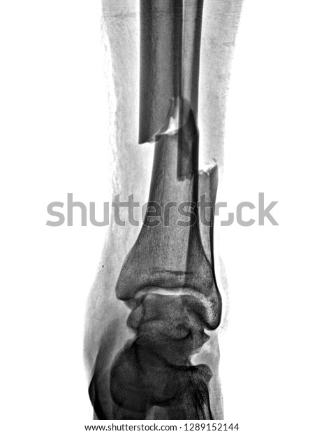
Cruris X Ray Anatomy Radiology Radiographic Foto Stok 1459919288 Shutterstock

Xray Rtankle Menemukan Lesi Osterolitik Intramedullary Dari Tibia Distal Kanan Foto Stok Unduh Gambar Sekarang Istock

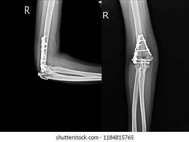
Xray Elbow Joint Aplateral Female Case Foto Stok 1184815765 Shutterstock

Ap Lateral Stok Foto Ap Lateral Gambar Bebas Royalti Depositphotos

Xray Image Ulnar Radius Fracture Ap Foto Stok 271398233 Shutterstock

Follow Dorso Palmar View Xray Distal Foto Stok 1724726155 Shutterstock

Fracture Distal Radius Colles Fracture Wrist Foto Stok 182503388 Shutterstock

Fraktur Siku Lengan Bawah Xray Gambar Menunjukkan Piring Dan Sekrup Fiksasi Foto Stok Unduh Gambar Sekarang Istock

Fraktur Konsolidasi Sinarx Tulang Kaki Bawah Foto Stok Unduh Gambar Sekarang Istock

Film Xray Pergelangan Tangan Menunjukkan Fraktur Distal Radius Foto Stok Unduh Gambar Sekarang Istock

